
MicroRNA Profiling in Mouse Testis Using Microarray Technology
Explore how microRNA microarray analysis reveals differential miRNA expression during mouse spermatogenesis and highlights regulatory mechanisms controlling germ cell development.
Introduction
Spermatogenesis is a highly coordinated biological process responsible for the production of mature male germ cells (spermatozoa). This process occurs in the seminiferous tubules of the testes and involves a complex sequence of cellular events including proliferation, meiosis, and differentiation of germ cells.
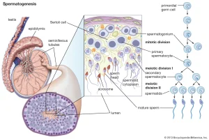
The regulation of spermatogenesis relies not only on transcriptional mechanisms but also on post-transcriptional regulatory processes that control mRNA stability and translation. Among the most important regulators involved in post-transcriptional control are microRNAs (miRNAs).
MicroRNAs are small non-coding RNA molecules, typically 19–23 nucleotides long, that regulate gene expression by binding to complementary sequences within the 3′ untranslated regions (3′-UTRs) of target messenger RNAs (mRNAs). This interaction results in translational repression or mRNA degradation, thereby modulating protein synthesis.
The first miRNA, lin-4, was discovered in the nematode Caenorhabditis elegans and was shown to regulate developmental timing. Since this discovery, hundreds of miRNAs have been identified across plants and animals, many of which are evolutionarily conserved.
Current studies suggest that miRNAs may regulate up to 30% of protein-coding genes in mammalian genomes. In addition, many miRNAs display strong tissue-specific expression patterns, highlighting their importance in specialized biological processes.
For example, miR-1 is predominantly expressed in cardiac and skeletal muscle tissues, indicating its role in muscle differentiation and function. Similar regulatory roles are expected in reproductive tissues.
Recent evidence suggests that miRNAs are involved in translational regulation during spermatogenesis. One example is miR-122a, which targets the transition protein 2 (Tnp2) gene. Tnp2 is a testis-specific gene involved in chromatin remodeling during sperm maturation.
Despite the increasing number of identified miRNAs, their expression patterns and regulatory roles in germ cells remain poorly characterized.
To better understand the involvement of miRNAs in spermatogenesis, researchers applied microarray-based miRNA profiling to analyze differences in miRNA expression between immature and mature mouse testes. The microarray results were further validated using quantitative real-time PCR (qRT-PCR), and computational analyses were used to predict potential target genes involved in germ cell development.
 Experimental Design and Biological Samples
The study was conducted using Balb/c male mice, which were maintained under standardized laboratory conditions.
Two developmental stages were investigated:
-
Immature mice: 1 week old
-
Mature mice: 7 weeks old
Each experimental group consisted of eight biological samples. Testicular tissues were collected immediately after euthanasia and preserved in liquid nitrogen to maintain RNA integrity.
All experimental procedures followed internationally accepted laboratory animal care guidelines.
RNA Extraction and Preparation
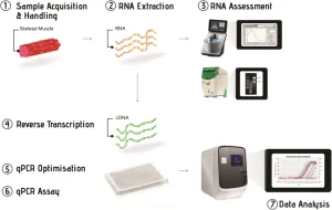
To analyze miRNA expression, total RNA was extracted from testicular tissue using the widely used TRIzol reagent protocol.
The procedure involved:
-
Grinding frozen testicular tissue into fine powder under liquid nitrogen.
-
Lysing the tissue using TRIzol reagent.
-
Isolating RNA through phase separation and precipitation.
-
Assessing RNA quality through gel electrophoresis.
High-quality RNA is essential for reliable microarray and PCR analyses, as degraded RNA may affect miRNA detection and quantification.
miRNA Microarray Analysis
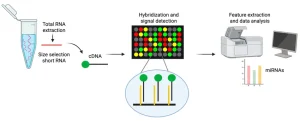
To investigate global miRNA expression patterns, researchers used miRNA microarray technology, which enables the simultaneous detection of hundreds of miRNAs.
The microarray platform used in this study contained:
-
509 oligonucleotide probes
-
Detection coverage for 892 miRNAs
-
miRNAs from human, mouse, and rat genomes
Each probe was printed three times on the array, improving measurement reliability and statistical confidence.
Microarray Workflow
The main steps included:
-
miRNA enrichment from total RNA using a specialized isolation kit.
-
Fluorescent labeling of miRNAs using Cy3 dye.
-
Hybridization of labeled miRNAs to the microarray probes.
-
Laser scanning of the microarray to measure fluorescence signals.
-
Digital image analysis to quantify miRNA expression levels.
To ensure experimental reproducibility, each microarray experiment was performed twice.
Data normalization and statistical analysis were performed using the Significance Analysis of Microarrays (SAM) software, which identifies significantly differentially expressed genes based on changes in expression relative to measurement variability.
Validation Using Quantitative Real-Time PCR
Although microarrays provide high-throughput expression profiling, quantitative real-time PCR (qRT-PCR) remains the gold standard for gene expression validation.
Because mature miRNAs are extremely short, conventional PCR amplification can be challenging. Therefore, the study used stem-loop reverse transcription primers, which improve specificity and sensitivity for miRNA detection.
qRT-PCR Workflow
-
Reverse transcription of RNA into cDNA using stem-loop primers.
-
Amplification of specific miRNA sequences using gene-specific primers.
-
Detection of PCR products using SYBR Green fluorescence.
-
Normalization using small nuclear RNA, a commonly used internal reference gene.
All PCR reactions were conducted in triplicate, including negative controls, to ensure accuracy.
Quantification of miRNA Expression
Relative miRNA expression levels were calculated using amplification efficiencies derived from standard cDNA dilution curves.
- This method allows accurate comparison of miRNA expression across biological samples.
Differential miRNA Expression During Spermatogenesis
Microarray analysis revealed significant differences in miRNA expression between immature and mature mouse testes.
Key Findings
-
19 miRNAs showed significant differential expression
-
14 miRNAs were upregulated in immature testes
-
5 miRNAs were downregulated
Among the upregulated miRNAs, three showed particularly strong expression increases:
-
mmu-miR-411
-
mmu-miR-495
-
mmu-miR-434-5p
These expression changes were confirmed by qRT-PCR, demonstrating strong agreement between the two analytical techniques.
Interestingly, some detected miRNAs corresponded to human or rat sequences without known mouse equivalents, suggesting the existence of previously uncharacterized mouse miRNAs.
Prediction of miRNA Target Genes
To better understand the biological significance of the differentially expressed miRNAs, researchers used computational target prediction tools, including:
-
TargetScan
-
PicTar
These bioinformatics algorithms identify potential miRNA binding sites in the 3′-UTR regions of mRNAs.
Several predicted target genes were associated with testicular development and germ cell differentiation, including:
-
Brd2 :involved in spermatocyte and spermatid development
-
Usp42 : expressed during embryogenesis and spermatogenesis
-
Rsbn1 : a transcription regulator in round spermatids
-
Edn1 : a signaling molecule active in mammalian testes
-
Sox5 and Sox6 : transcription factors regulating gene expression
-
NR6A1 (GCNF) : involved in transcriptional control during meiosis
These predicted interactions highlight the complex regulatory networks controlled by miRNAs during germ cell development.
Biological Implications
Spermatogenesis is a multistep developmental process that transforms diploid germ cells into haploid spermatozoa through tightly regulated stages.
The findings of this study indicate that miRNAs play a critical regulatory role during early spermatogenic development, particularly in immature testicular tissue.
The higher expression levels of several miRNAs in immature testes suggest that these molecules may regulate gene expression programs required for germ cell differentiation and maturation.
Furthermore, the predicted target genes identified in this study are involved in chromatin remodeling, transcriptional regulation, and cellular differentiation, all essential processes for successful spermatogenesis.
Conclusion
MicroRNA profiling using microarray technology combined with qRT-PCR validation provides valuable insights into the molecular mechanisms regulating spermatogenesis.
This study identified 19 miRNAs that are differentially expressed between immature and mature mouse testes, suggesting that miRNAs play an important role in controlling gene expression during germ cell development.
The predicted target genes further support the hypothesis that miRNA-mediated regulation contributes to the complex genetic networks underlying spermatogenesis.
Future studies focusing on the functional characterization of these miRNAs will provide deeper insights into male reproductive biology and fertility regulation.